oral diagnosis
Medical history, General physical examination and Oral examination

A thorough history is taken before the examination regarding duration and frequency of tobacco use in any form like cigarette, beedi, chewing pan, gutka, khaini etc and of alcohol consumption.
Oral examination: A careful examination of entire inner cavity of the mouth which includes the roof of mouth, back of the throat, and inside of cheeks and lips is then carried out. The doctor looks for red or white patches or any other abnormal areas over head, neck or face. He/she also examines for any lumps, swelling or any other problem with the nerves of mouth or face. If any abnormal area is found during examination, it is confirmed by further tests which are detailed below.
Invasive Tests
Brush cytology: In this test, the suspected area/lesion is brushed and the cells are looked at under microscope for abnormal cells by a pathologist.- NFine Needle Aspiration Cytology (FNAC): FNAC is generally used to diagnose metastatic carcinoma of head and neck, in the cervical region. In this test, a thin needle which is attached to a syringe is used to draw few cells from the suspected lump or swelling. These cells are smeared onto a glass slide, then stained and examined under microscope by a pathologist to examine for abnormal cells.
- NBiopsy: A small piece of tissue is taken from suspicious area using a punch biopsy instrument. Sometimes it may be done under the guidance of endoscopy, if the lesion is not easily accessible. This tissue is processed in the laboratory and examined for presence or absence of cancer.
Imaging Tests.
Imaging tests are done to confirm the diagnosis, document the extent of spread of disease, staging etc. The most common diagnostic imaging tests are X-rays, CT scan, MRI and PET scan.Other Tests.
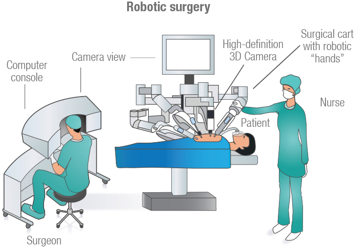
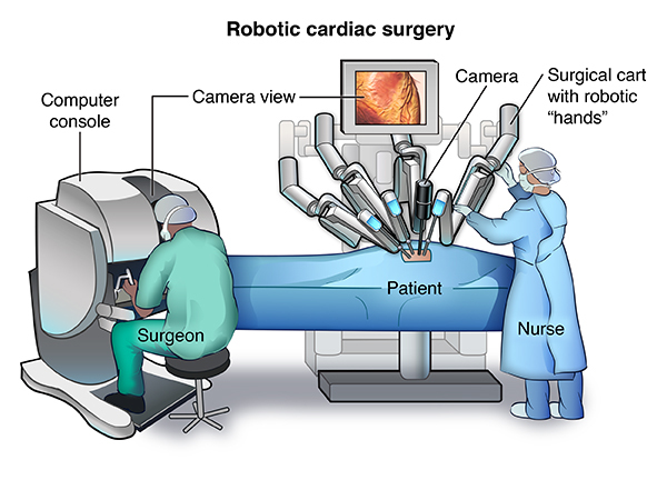
Human Papillomavirus (HPV) Testing: Oral cancers with HPV infection are on the rise. Doctors may test the biopsy sample for the presence of HPV infection as the possible cause.During a da Vinci robotic surgery procedure, the surgeon makes a few tiny incisions to insert miniaturized instruments and a high-definition camera inside the patient. The camera allows the surgeon to view a highly magnified, high-resolution 3-D image of the surgical site.
Seated comfortably at an ergonomically designed console, with eyes and hands in line with the instruments, the surgeon uses controls below the viewer to move the instrument arms and camera. The da Vinci Surgical System then translates, in real time, the surgeon’s hand, wrist and finger movements into precise movements of the instruments inside the patient.
Throughout a da Vinci robotic surgery procedure, the surgeon controls every surgical maneuver. The system cannot be programmed or act in any way without the surgeon’s input.
